The spine is a very complex mechanical structure that is highly flexible yet very strong and stable. In the normal spine, regardless of your position or activity, including sleeping, there is always some type of physical demand being placed on it.
The primary functions of the spine include:
- Protect the spinal cord, nerve roots, and internal organs
- Provide flexibility of motion
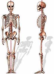
- Provide structural support and balance for upright posture. The spine bears the load of the head, shoulders and arms, and upper body. The upper body weight is then distributed to the hips and legs. The spine attempts to keep the body’s weight balanced evenly over the pelvis. This reduces the amount of work required by the spinal muscles and can eliminate muscle fatigue and back pain.

 |
 |
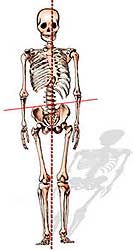 |
Loss of spinal balance can result in strain to the spinal muscles and deformity of the spine as it attempts to maintain an upright posture.
Regions of the Spine
 There are 33 vertebrae (bones) in the spine. Anatomically, the spine is divided into four regions:
- The top 7 vertebrae that form the neck are called the cervical spine and are labeled C1-C7.
- The upper back, or thoracic spine, has 12 vertebrae, labeled T1-T12.
- The lower back, or lumbar spine has 5 vertebrae, labeled L1-L5.
- The sacrum and coccyx (tailbone) are made up of 9 vertebrae that are fused together to form a solid bone. The sacrum is labeled S1.
Curves of the Spine
When viewed from the front or back, the normal spine is in a straight line, with each vertebra sitting directly on top of the other. A side-to-side curve in the spine is called a scoliosis.
When viewed from the side, the normal spine has three gradual curves:
- The neck has a lordosis; it curves towards the back.
- The thoracic spine has a kyphosis; it curves towards the front.
- The lumbar spine also has a lordosis.
These curves help the spine to support the load of the head and upper body, and maintain balance in the upright position.
Vertebrae
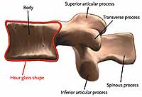
Although the vertebrae have slightly different appearances as they range from the cervical spine to the lumbar spine, they all have the same basic structures, and the structures have the same names. Only the first and second cervical vertebrae are structurally different in order to support the skull.

 |
 |
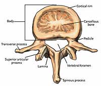
The anterior arch is called the vertebral body. Discs connect one vertebral body to another to allow motion of the spine and cushion it against heavy loads. Together, the vertebral bodies and discs bear about 80 percent of the load to the spine.
 |
 |
The posterior arch consists of the pedicles, laminae, and processes.
The pedicles are two short cylinders of bone that extend from the vertebral body. Nerve roots branch off the spinal cord and exit to the body between the pedicles of two vertebrae. If the spine becomes unstable, the pedicles may compress the nerve root, cause pain or numbness.
Laminae are two flattened plates of bone that form the walls of the posterior arch. Over time, the laminae may thicken, a process called stenosis. This thickening compresses the spinal cord and/or nerves causing pain or numbness.
The articular, transverse, and spinous processes project off the laminae. Ligaments and tendons attach to the processes. The articular processes join one vertebra to another posteriorly.
The transverse processes extend out on either side of the laminae. The spinous process is the bony projection that can be felt through the back of someone’s skin.
Intervertebral Discs
Intervertebral discs are located between each vertebra from C2-C3 to L5-S1. Combined, they make up one fourth the height of the spinal column. The discs act as shock absorbers to the loads placed on the spine and allow movement of the spine. Movement at a single disc level is limited, but all of the vertebrae and discs combined allow for a significant range of motion.
 |
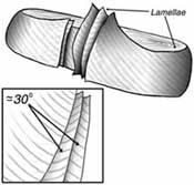 |
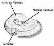
The intervertebral disc is made up of two components: the annulus fibrosus and the nucleus pulposus. The annulus fibrosus is the outer portion of the disc. It is composed of layers of collagen and proteins, called lamellae. The fibers of the lamellae slant at 30-degree angles, and the fibers of each lamella run in a direction opposite the adjacent layers. This creates a structure that is exceptionally strong, yet extremely flexible.
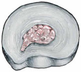
The nucleus pulposus is the inner gel material surrounded by the annulus fibrosus. It makes up about 40 percent of the disc. This ball-like gel is contained within the lamellae. The nucleus is composed primarily of loose collagen fibers, water, and proteins. The water content of the nucleus is about 90 percent at birth and decreases to about 70 percent by the fifth decade.
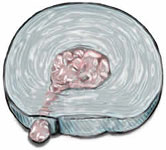
Injury or aging of the annulus fibrosus may allow the nucleus pulposus to be squeezed through the annulus fibers either partially, causing the disc to bulge, or completely, allowing the disc material to escape the disc. The bulging disc or nucleus material may compress the nerves or spinal cord, causing pain.
 |
 |
In the early years of life, the discs have a blood supply that nourishes them. In the second and third decades, discs gradually lose this blood supply, until they are avascular. At this point, the disc begins to degenerate, or age. By the age of 50, over 95 percent of all people will have disc degeneration. The disc begins to lose water content and shrinks. The spine’s range of motion and shock-absorbing ability are decreased. This may result in injury to the nerves and vertebrae, and the aging disc itself may generate pain.

Spinal Cord and Nerve Roots
 The brain and spinal cord together make up the central nervous system. The spinal cord is located immediately below the brain stem. It extends through the foramen magnum, a hole at the base of the skull.
The spinal cord functions as a sophisticated network that carries information from the outer elements of the body (skin, muscles, ligaments, joints) through the sensory tracts, to the central “computer,” the brain. Data are processed there, and new information such as muscle control is sent out through the motor tracts of the spinal cord.
The spinal cord ends as the conus medullaris at the L1 vertebral level, where it branches into the cauda equina, a collection of nerves that extend from the conus medularis to the sacrum. The conus medularis nerves float freely in spinal fluid, making it possible to pass a needle safely into the area to draw a sample of spinal fluid or inject drugs, anesthetics, or radiologic substances for x-ray, MRI or CT scan.